
Microglia: Brain macrophages
Microglia is better known as the brain’s resident immune system, or brain macrophages. Microglia regulation of neuron activity was missed until very recently. It is only since 2009 that the multifaceted nature of microglia involvement in neuron signaling and plasticity of brain synapses also became recognized. Microglia is one of several types of cells occupying the brain and spinal cord.
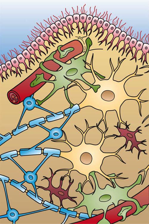
Brain cells, Holly Fischer/Wikimedia Commons
This image shows two blood capillaries and the four different types of cells found in the brain: Ependymal cells (light pink), Astrocytes (green), Microglia (dark red), and Oligodendrocytes, which are functionally similar to Schwann cells in the spinal cord (light blue).
Origin of microglial cells
Microglia is of embryonic mesodermal origin. It appears very early in embryological development during primitive blood cell formation in the yolk sac. This is long before bone marrow blood cells appear. Thus, microglia and bone-marrow-originated macrophages represent two genetically distinct populations.
Unlike peripheral immune system macrophages that are short lived, microglia is long-lived and subject to the influences of normal aging. Central nervous system resident times for microglia extend across much of an animal’s lifespan. This contrasts with the few hours to few days that peripheral macrophages survive.
Brain microglia
Brain microglia, prior to pathogen exposure, possesses a very small cell body with many long-branched processes extending into the surrounding area. The processes continuously contact and elongate to scan the tissue environment. This form of microglia is named surveying microglia.
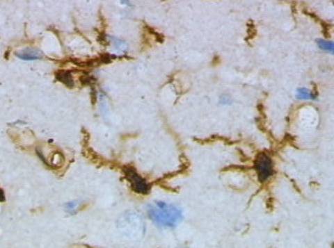
Brain Tissue Stained to Display Microglia, Grzegarz Wicker/Wikimedia Commons
Activated microglia
One aspect of microglia function is to act as a pathogen sensor. Microglia becomes ‘activated’ upon infection with bacteria and viruses. In its activated macrophage state, microglia can engulf and digest infectious particles and damaged cellular material.
When infectious agents enter the brain, activated microglia may recruit T cells and macrophages across the blood-brain barrier to limit damage.
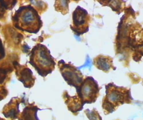
Activated microglia are brain macrophages, Grzegarz Wicker/Wikimedia Commons
In the adult, microglia represent about 10% of the cells in the brain and spinal cord. It is evenly distributed through all brain regions. Processes of neighboring microglial cells do not overlap, and each microglia cell acts as a scavenger for its immediate area.
Sensors – proteins also known as receptors – in the membrane of microglia processes detect signals (chemicals called ligand) requiring a response from microglia. When it is a pathogen signal that is detected, microglia processes retract, and the cell body becomes mobile. Mobile, activated microglia advances toward the signal and becomes a phagocytic microglia.
Surveying microglia also migrates to sites of non-infectious tissue injury. It is drawn by local tissue signaling molecules to the damaged area. At the site of injury, it evaluates the extent of the damage. If the problem is repairable, microglia mends injured cells by secreting growth factors. If the damage is too great and the cell is dying, microglia become activated, finishes the kill and clears the damage.
Neuron synapse repair
Microglia oversees neuron repair. There are a multitude of receptor proteins on microglia cell membrane. Ligands that bind those receptors, and thereby initiate response of microglia, may originate from pathogens, dying tissue, or be secreted by healthy neurons, oligodendrocytes or astrocytes.
Only subsets of ligands for microglia receptors mobilize and activate microglia turning it into an immune responder. Some signals in the normal brain environment have the opposite effect of maintaining the characteristics of surveying microglia.
Surveying processes of microglia make brief about 5-minute, direct contact with brain synapses at a frequency of about once per hour. Such contact increases with rising neuron activity and decreases when there is less synaptic activity.
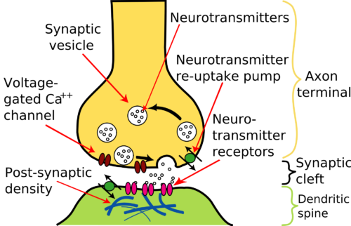
Basic elements of a brain synapse, Nrets/WikiMedia Commons
Active synapses release ATP into the extracellular environment and it is the level of ATP that is being monitored by microglia surveying processes. ATP is a chemo-attractant that induces microglia to polarize its processes toward active synapses.
The frequency of contact at neuron synapses by microglia processes is interpreted to suggest that microglia diligently monitors and responds to the functional status of synapses. Data obtained with live imaging studies are consistent with a theory that the duration of microglia contact is directly related to which synapses are pruned in the process of neuron remodeling.
Dendritic spines where the duration of contact by microglia is extended are more often eliminated than those where the contact is short. In this image, an early drawing by Cajal, dendritic spines are visible on the dendrites of hippocampus neurons.
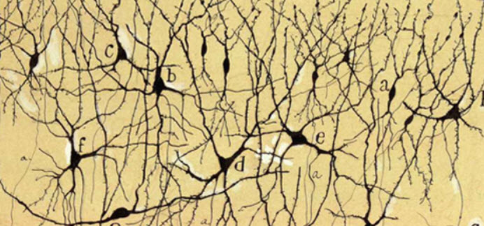
Neurons of the hippocampus showing dendritic spines, Cajal Public Domain/Wikimedia Commons
The capacity for microglia to phagocytose synapses is currently being evaluated. But, the dynamic behavior of microglia processes at neuron synapses seems to be non-random. Unnecessary synapses appear to be tagged for pruning by deposition of complement proteins. It is likely that several steps are involved in the pruning process.
First, the level of synaptic activity is determined, then alterations in nerve synapse protein glycosylation is monitored, then tagging of selected synapses with complement proteins is accomplished, and finally dendritic spines are removed by microglia processes in a phagocytic manner.
Aging brain
Microglia may change its function in the aging brain. Microglia is thought to play a variety of roles in most neurodegenerative diseases including Alzheimer’s, Parkinson, and Huntington’s. One common factor in these diseases of the aging brain is the lack of physiologic regulation of the brain’s major excitation neurotransmitter, glutamate.
Excess extracellular glutamate is toxic to neurons. It is recognized that microglia activated by pathogen or brain damage secretes glutamate using a membrane exchange transporter. Under conditions of mild activation other cells, astrocytes, can scavenge the excess glutamate. In diseased states such as Alzheimer’s activated microglia is so extensive that astrocytes are unable to maintain tissue glutamate at normal physiologic levels.
More about the role of microglia in the neurodegeneration observed in Alzheimer’s disease can be found in my book ‘Inside the Closed World of the Brain, How brain cells connect, share and disengage – and why this holds the key to Alzheimer’s disease.” To take a look inside this book click here.
Considering brain microglia as a regulatory partner in neuron activity in healthy brain in addition to its role as brain macrophage, paves the way for creating more effective methods of preventing neurodegenerative diseases of aging brain. It is expected that ongoing research will continue to expand our knowledge of microglia cell function within the next few years.
Do you have questions?
Please put your questions in the comment box or send them to me by email at DrReece@MedicalScienceNavigator.com. I read and reply to all comments and email.
If you find this article helpful share it with your fellow students or send it to your favorite social media site by clicking on one of the buttons.
Further reading
Astrocytes and Energy in the Brain

Margaret Thompson Reece PhD, physiologist, former Senior Scientist and Laboratory Director at academic medical centers in California, New York and Massachusetts is now Manager at Reece Biomedical Consulting LLC.
She taught physiology for over 30 years to undergraduate and graduate students, at two- and four-year colleges, in the classroom and in the research laboratory. Her books “Physiology: Custom-Designed Chemistry”, “Inside the Closed World of the Brain”, and her online course “30-Day Challenge: Craft Your Plan for Learning Physiology”, and “Busy Student’s Anatomy & Physiology Study Journal” are created for those planning a career in healthcare. More about her books is available at https://www.amazon.com/author/margaretreece. You may contact Dr. Reece at DrReece@MedicalScienceNavigator.com, or on LinkedIn.
Dr. Reece offers a free 30 minute “how-to-get-started” phone conference to students struggling with human anatomy and physiology. Schedule an appointment by email at DrReece@MedicalScienceNavigator.com.