 A Virtual Tour
Inside the Closed World of the Human Brain
![]() Welcome to a week-long tour of human brain physiology. This tour is in partnership with The Dana Alliance for Brain Initiatives during their Brain Awareness Week Campaign.
Welcome to a week-long tour of human brain physiology. This tour is in partnership with The Dana Alliance for Brain Initiatives during their Brain Awareness Week Campaign.
Founded in 1996, Brain Awareness Week unites the efforts of partner organizations from around the world in a week-long celebration of the brain every March.
These 7 daily tour topics seek to discover the secret of those who live past 100 years and still retain their mental competence.
If you are struggling to understand all the talk in the media about the human brain and dementia, then do not miss this read. Before a thing that is broken can be fixed, there is a need to know how it is supposed to work.
To uncover the facts about dementia, begin by learning how a healthy human brain operates.
Read more about the author of this tour inside the closed world of the brain Dr. Margaret Reece at amazon.com/author/margaretreece
Brain’s Scientific Language
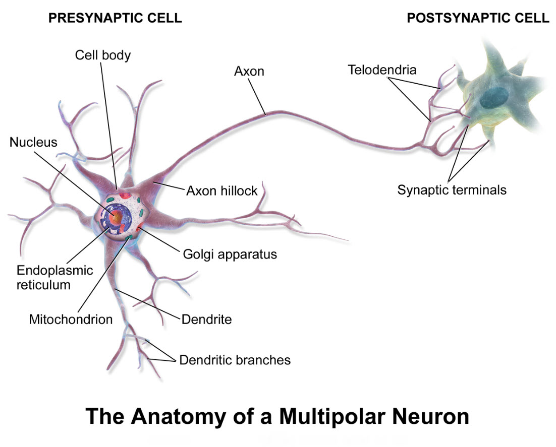
Neuron anatomy, BruceBlaus/Wikimedia Commons
The strange scientific language used to describe the brain and its population of cells makes it difficult to follow scientific discussions of this subject.
Scientific labels persisting in medicine are in Classical Latin. Classical Latin was the universal language of large segments of the western scientific world from the time of the Roman Empire through the 17th century.
Psychologists now tell us that words, even strange words, are learned fast by the human brain when they are associated with something familiar.
Read about how to connect medical terms describing brain function with familiar things you already know by clicking here.
How the Human Brain Is Organized
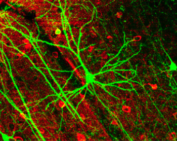
Cortical pyramidal cell capture by confocal microscopy. Photomicrograph from Lee WCA Plos Biol 4(2):E29 (2006)/Wikimedia Commons
Brain structure is described in three ways. First, visual observation of the whole brain establishes the overall layout of larger structures.
Second, microscopic visualization of fixed, sliced and stained brain tissue reveals its cellular structure.
Third, videos of living brain obtained with computerized microscopes demonstrate mobility of resident cells.
Neuron signaling practices in the human brain are more complex than those of other species. Yet, the brain’s visual structures are similar among mammalian species.
And, much of what is known about the human brain’s cellular operational systems comes from observations of rats, mice and non-human primates.
Learn more about the arrangement of the densely-crowded occupants of the human brain by clicking here.
Quality Control Of Brain’s Watery Environment
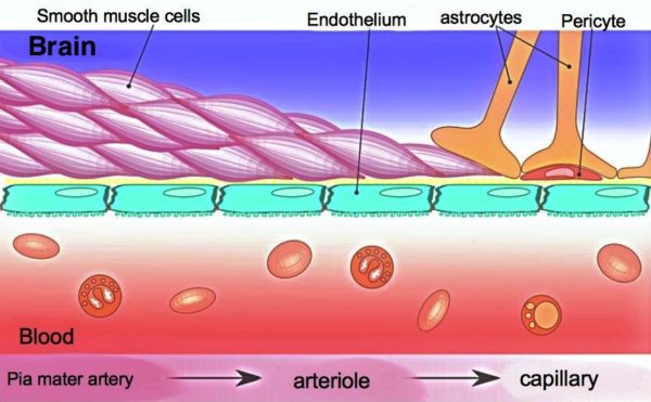
Brain Capillaries are reinforced by pericytes and astrocytes, Armin Kubelbeck/Wikimedia Commons
Neurons of the brain are ‘electrical’ cells. A consistently precise chemical composition of the brain fluid surrounding neurons is essential for their electricity.
The brain’s quality control program for its watery fluids includes strict restriction of entry for most substances in blood plus a circulatory system with no comparison elsewhere in the body, the cerebrovascular circuit.
Read more about the blood brain barrier and other circulatory adaptations isolating the brain from wide fluctuations in blood nutrients created by feasting and fasting by clicking here.
Neurons & Brain’s Other 90% Of Cells
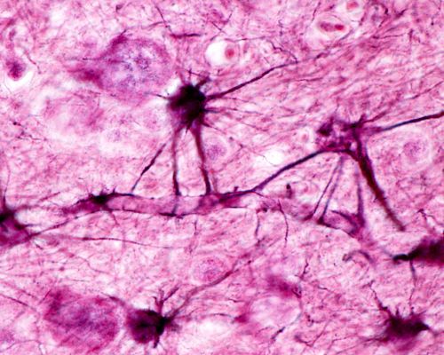
Astrocytes along a blood vessel in the brain, Jose Luis Calvo/Shutterstock.com
Only 10% of brain’s cells are neurons. The remaining 90% of brain cells, called glia and microglia, participate as intimate partners of the neurons in management of brain function.
There are two types of glia, astrocytes and oligodendrocytes. Both astrocytes and oligodendrocytes mature out of the same stem cell population as neurons.
Microglia, the resident immune system of the brain originates during early embryo development from the extra-embryonic yolk sac. Microglia of the yolk sac migrates into neural tissue before bone and bone’s associated immune cells develop. Immune cells of bone origin seldom enter the brain.
Brains of people with Alzheimer’s dementia and Parkinson’s disease display a disruption of the normal supportive relationship between neurons, astrocytes, oligodendrocytes and microglia.
Read a description of the interdependence of the brain cell community by clicking here.
Modern Brain Imaging Research
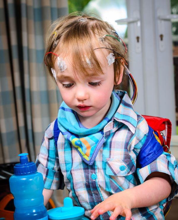
Boy with EEG electrodes, Steve Buckley/Shutterstock.com
Brain imaging has greatly increased understanding of brain function. Many theories stemming from early brain anatomic studies speculated that variation in neuron structure, neuron location in the brain, and how neurons connected with each other regulated distinct characteristics of brain performance.
Early confirmation that intellectual pursuits align with discrete regions of the human brain emerged from examination of patients in the clinic who suffered local brain damage.
Modern brain studies apply non-invasive methods to explore information handling by different parts of the brain. Five neuron imaging techniques dominate this research.
They are event Related Potentials (ERPs) of EEG, Magnetic Resonance Imaging (MRI), Functional Magnetic Resonance Imaging (fMRI), Magnetoencephalography (MEG) and Functional Near Infrared Spectroscopy (fNIRS).
For how each of these imaging techniques provides different information about brain performance click here.
Human Memory Formation & Recall

Remember the cat helping us study? Dmitry Melnikov/Shutterstock.com
Most systems for cataloging the defining characteristics of human memory originated in the field of psychology. One type of memory described by psychologists is episodic memory. Episodic memory recalls specific events, situations and personal experiences.
Studies of episodic memory over the past 15 years by neuroscientists produced the clearest and best accepted data about how memory is managed in the human brain.
Episodic memory is first coded by neurons of the only part of the human brain that can produce new neurons from neural stem cells late into old age.
Fragments of a coded memory scatter to multiple separate brain locations for storage. Visual representation, auditory representation, and the emotional component of an event may all map to different brain areas.
Find out more about how fragments of a memory are recalled and updated with information obtained since the event occurred by clicking here.
Dissecting Alzheimer’s Dementia
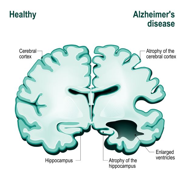
Illustration of what Alzheimer’s does to the human brain, Designua/Shutterstock.com
What exactly goes awry in Alzheimer’s disease? Recent studies reveal that Alzheimer’s dementia is a slow dying of neurons in regions of the brain needed for episodic memory of people and events.
Those who develop Alzheimer’s dementia lose their ability to remember and reason in a progression that is slow, frustrating and ultimately fatal. There are measurable changes in memory forming brain regions for decades before symptoms appear.
The brain imaging techniques described two days ago on this blog tour are helping scientists unravel the cellular behaviors that lead to neuron death and memory loss.
Yet, a definite diagnosis of Alzheimer’s dementia is obtained only after death with microscopic examination of brain tissue. A critical element missing in studies of Alzheimer’s dementia before 1997 was the lack of a control group, long-lived individuals who had maintained mental competence until their death and were willing to donate their brain to science.
Discover how the brain tissue now becoming available from long-lived, mentally competent people compare with that of young healthy brain and the brains of Alzheimer’s sufferers by clicking here.
Margaret Thompson Reece PhD, physiologist, former Senior Scientist and Laboratory Director at academic medical centers in California, New York and Massachusetts is now Manager at Reece Biomedical Consulting LLC.
She taught physiology for over 30 years to undergraduate and graduate students, at two- and four-year colleges, in the classroom and in the research laboratory. Her books “Physiology: Custom-Designed Chemistry”, “Inside the Closed World of the Brain”, and her online course “30-Day Challenge: Craft Your Plan for Learning Physiology”, and “Busy Student’s Anatomy & Physiology Study Journal” are created for those planning a career in healthcare. More about her books is available at https://www.amazon.com/author/margaretreece. You may contact Dr. Reece at DrReece@MedicalScienceNavigator.com, or on LinkedIn
Featured header image: ©Moonlight Photo Studio/Shutterstock.com
Reece Biomedical Consulting LLC Privacy Policy: Click HERE