
Article #2 of a 7 Part Series
Brain structure is described in three ways.
First is its gross anatomy, its large characteristics that are visible to the eye.
Second is its cellular structure, visible with the help of a classic light microscope.
Third is its dynamic cellular movement, captured by computerized systems that convert fluorescent signals from the brain into images and videos.
Human Brain Anatomy
Brains are very soft, gel-like structures. To protect their fragile makeup, brains are enclosed within a bony case, the skull. The only part of human brain anatomy not under bone is the eyes. Unlike other sensory organs that send their signals to the brain via their neuron axons, the eyes are brain tissue.
The term gross anatomy refers to the external features of an organ that can be observed visually and by feel.
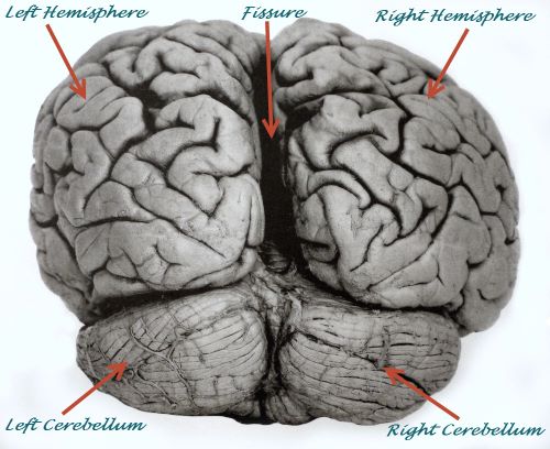
Human brain showing left and right hemispheres, cerebellum and central fissure, Pete Spiro/Shutterstock.com
The gross organization of the brain is similar among mammalian species. There are two large pieces called the right and left hemispheres.
The hemispheres are connected to each other by a bridge of neuron axons named the corpus callosum.
Within this axon bridge, information travels in both directions functionally connecting the hemispheres.
Corpus callosum comes from two Latin words with corpus referring to a body of tissue and callosum indicating its hard texture, much like the consistency of a callus.
The axons of the corpus callosum connect corresponding parts of the hemispheres and permit the right and left hemisphere to coordinate their activity.
The surface of the brain hemispheres is deeply folded tissue. The top of a ridge is named gyrus and the trough is named sulcus. Gyrus is a Latin word meaning a circular or spiral form. Whereas, sulcus is a derivative of the Latin word sulcätus, which means to furrow.
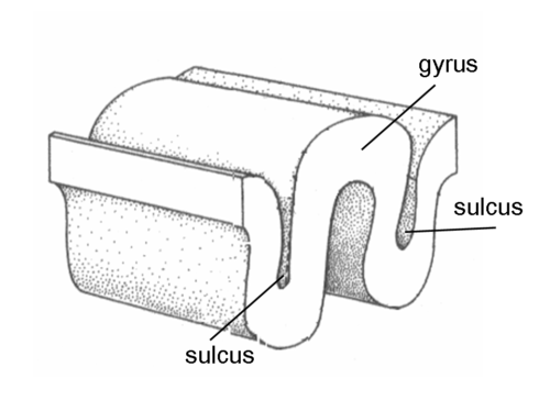
Illustration of a gyrus vs. a sulcus, Public Domain Image/Wikimedia Commons
There are two additional large structures behind the hemispheres named the right and left cerebellum. The cerebellum is connected to the brain hemispheres by a small structure named the pons. In Latin the word pons means bridge and cerebellum is a diminutive form of cerebrum, the Latin word for brain. Put together the name of this part of the brain, the part that coordinates complex movement, posture and balance translates into little brain.
Neuron Cell Structure
Neuron cell structure was not observed until the end of the 19th century. Because of the brain’s soft gel-like appearance, it was considered an irrelevant organ for millennia. Even through the first half of the 1800s scientists claimed that brain was the only organ without cells.
Normal histology stains used at the time to distinguish cellular structure of other tissue did not work in the brain, because the chemicals were widely absorbed by fatty material distributed throughout the brain. In 1873, Camillo Golgi developed a silver process capable of staining brain neurons. It turned out that Golgi’s silver stain is only taken up by a small percentage, 1% to 3%, of neurons in a tissue slice.
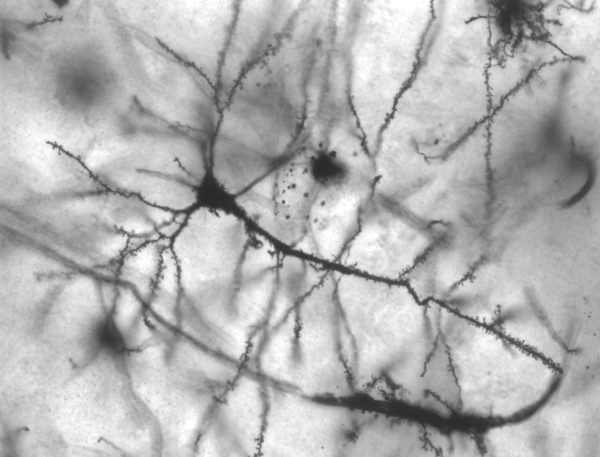
Pyramidal hippocampus neuron showing dendritic spines/MethoxyRoxy/Wikimedia Commons
This is fortunate however because a stain that marks all of the neurons would obscure their structure. With an estimated 86 billion neurons and about 10 times as many partnering cells plus 15-25 square meters of blood vessels, space in the brain is limited.
Santiago Ramón y Cajal and Camillo Golgi shared the 1906 Nobel Prize for their extensive body of work describing the structure of brain neurons. Santiago Ramón y Cajal proposed neurons may be separate entities that communicate with each other across narrow spaces. Other scientists, including Camillo Golgi, did not embrace the idea at first. But, Santiago Ramón y Cajal was correct.
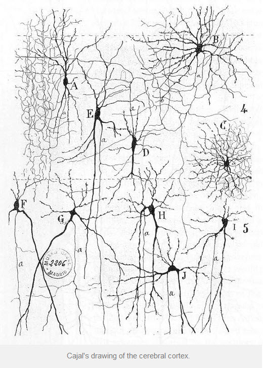
Drawing of Neurons of Cerebral Cortex, Ramon y Cajal, Public Domain Image/Wikimedia Commons
The structures at the places where neurons connect with each other are collectively called synapses. Synapse derives from a Greek work describing a point of contact.
Brain synapses occur at multiple places on neurons, at dendrites, cell bodies, axons and axon terminals.
The complex placement of neuron synapses in the brain permits fine-tuned regulatory control of neuron input and output and creates complex neuronal circuits that act as units.
Optical Imaging
A method of microscopy developed since 1990, optical imaging allows scientists to study living brains. This procedure employs instruments known as optical imaging systems. This is an invasive procedure restricted to use in animal studies. It requires introduction of genes to fertilized ova to produce fluorescing molecules in the neurons of the brain. It also requires thinning of the bone of the skull, or skull removal over the area of interest.
Fluorescence induced by optical imaging systems in brain tissue is too low to be detected by the human eye. The con-focal and 2-photon imaging microscopes that detect brain fluorescence are not microscopes in the same sense as traditional light microscopes. Instead, fluorescent light information is analyzed by the computerized optical imaging devices and a computer-created picture displays the information in a form the human eye can recognize.
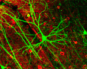
Cortical pyramidal cell capture by confocal microscopy. Photomicrograph from Lee WCA Plos Biol 4(2):E29 (2006)/Wikimedia Commons
Computerized microscopic techniques not only permit observation of brain anatomy in greater detail but also display the animated nature of brain tissue. Videos showing neuron animation captured with optical imaging systems are available on YouTube. Two of these are Actin in Dendritic Spines by Michael J Shell and
Neuron Time Lapse Video by Leigh Needleman
![]() Click Here for More about “How Does the Brain Develop?” – The Dana Alliance for Brian Initiatives
Click Here for More about “How Does the Brain Develop?” – The Dana Alliance for Brian Initiatives

Margaret Thompson Reece PhD, physiologist, former Senior Scientist and Laboratory Director at academic medical centers in California, New York and Massachusetts is now Manager at Reece Biomedical Consulting LLC.
She taught physiology for over 30 years to undergraduate and graduate students, at two- and four-year colleges, in the classroom and in the research laboratory. Her books “Physiology: Custom-Designed Chemistry”, “Inside the Closed World of the Brain”, and her online course “30-Day Challenge: Craft Your Plan for Learning Physiology”, and “Busy Student’s Anatomy & Physiology Study Journal” are created for those planning a career in healthcare. More about her books is available at https://www.amazon.com/author/margaretreece. You may contact Dr. Reece at DrReece@MedicalScienceNavigator.com, or on LinkedIn
Reece Biomedical Consulting LLC Privacy Policy: Click HERE
Featured header image: ©Moonlight Photo Studio, via Shutterstock.com