
Autonomic control of the body’s smooth muscle
No description of basic human physiology is complete without talking about the autonomic nervous system’s control of the body’s smooth muscle.
Location of smooth muscle
This illustration of a vascular bundle points out the smooth muscle of arteries and veins. Notice that the artery has a thick band of smooth muscle, and the vein has little smooth muscle.
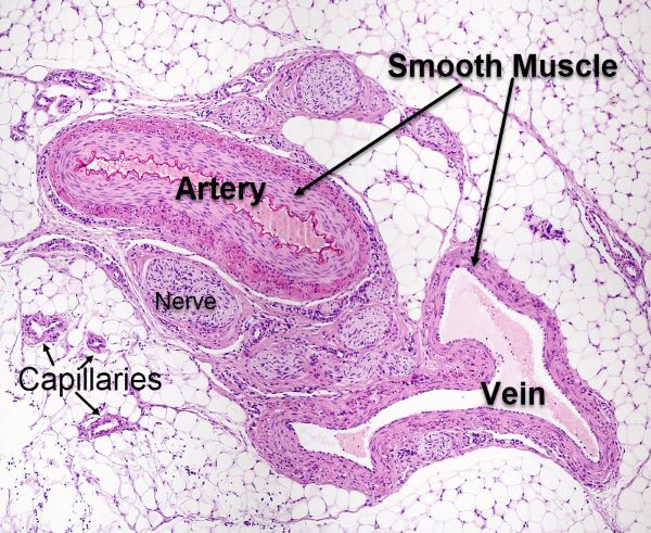
Comparison of smooth muscle presence in artery and vein, Jose Luis Calvo, Shutterstock.com
Smooth muscle of the veins help move blood back to the heart. The thick arterial smooth muscle is important to maintaining blood pressure between heart beats and diversion of blood to the skeletal muscles during exercise.
Smooth muscle is also found in the walls of hollow organs like the stomach, intestines, urinary bladder, and in the walls of passageways like those of the respiratory, urinary, and reproductive systems.
Smooth muscles are even found in the eyes around the iris and lens. In skin, smooth muscle causes hair follicles to erect in response to cold temperature. Smooth muscle forms the soft tissue of the internal organs of the body, the organs of the thoracic and abdominal cavities.
Smooth muscle contraction
Smooth muscle contraction occurs without our being aware of it. These muscle cells are not striated like skeletal and cardiac muscle. Rather, they have an arrangement of contractile proteins better suited to their shape. Smooth muscle are cells are shaped like spindles. They have tapered ends and are wide in the middle.
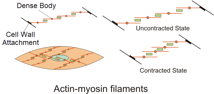
Actin and myosin filaments of smooth muscle, Boumphreyfr/Wiki Media Commons
The actin and myosin contractile apparatus is arranged in a net-like pattern in smooth muscle cells. Rather than lying in parallel, the actin-myosin filaments run diagonal to each other. At either end of the contractile chain, the actin molecules are attached to the cell plasma membrane forming dense bodies. When smooth muscle contracts the actin-myosin intermediate filaments shorten between the dense bodies. This shortens the long thin spindle-shaped cells, and they become fatter.
Smooth Muscle Motor Unit
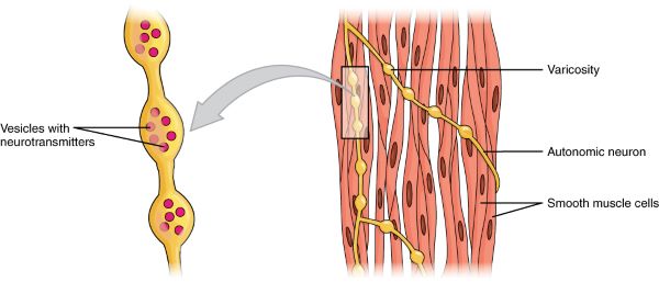
Design of smooth muscle motor unit, Open Stax, Wiki Media Commons
Sheets of smooth muscle cells contract as bundles. As illustrated here, smooth muscle motor units are arranged differently than skeletal muscle motor units.
Smooth muscle motor units are created by the terminal end of an autonomic motor neuron wrapping around a group of smooth muscle cells. Varicosities along the neuron terminus contain vesicles filled with neurotransmitter.
Action potentials reaching the autonomic neuron terminus release neurotransmitter at the points of contact between the varicosities and the smooth muscle cells. Neurotransmitter spreads over the nearby muscle cells, which contract as a unit.
Autonomic nervous system
Smooth muscle contraction is regulated by the brain through the neurons of the autonomic nervous system. Here is an illustration the motor nerves of the autonomic nervous system that cause smooth muscle contraction and relaxation. I know this slide is busy, but I will walk you through its major points.
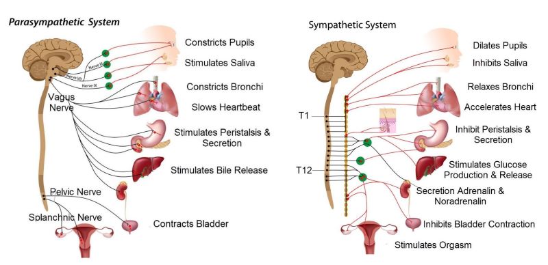
Both divisions of the autonomic system innervate each organ, Alila Medical Media, Shutterstock.com
First, the lines going from the brain and spinal cord are nerves. Remember the nerves are bundles of neuron axons. The cell bodies of the neurons included in these nerves reside in the brain stem, spinal cord, and spinal ganglion. I will get to the spinal ganglion shortly.
What I want you to notice in this slide is that there are two parts to the autonomic nervous system. They are the parasympathetic division and the sympathetic division. If you are wondering why I want you to know this, it is because you will need this information later to understand the physiology of exercise and of the fight-or-flight response.
Both autonomic divisions send nerves to the same visceral organs. They do that because they have opposing effects at each organ. For example, the parasympathetic system constricts the bronchi of the lungs and slows beating of the heart when the body is at rest. In contrast, the sympathetic system opens the bronchi and accelerates heartbeat during exercise.
The two autonomic nervous system divisions are reciprocally active. That is, when parasympathetic activity is high, sympathetic activity is always low. And the reverse: when sympathetic activity is high, parasympathetic activity is always low.
Insular Cortex
The cortical neurons of the brain control which autonomic division dominates smooth muscle contraction.
Sympathetic and parasympathetic nerves often carry axons of both smooth muscle motor neurons to the visceral organs and sensory neurons to the brain. the sensory cortex for information coming from the viscera is called the insular cortex. The insular cortex lies deep in the lateral sulcus on each side of the brain.
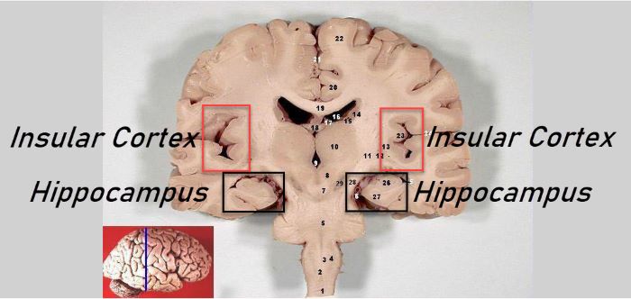
Location of the human insular cortex and hippocampus, John A Beal, Wiki Media Commons
Combined activity of the motor neurons of the insular cortex of the brain (outlined by the red boxes), the hypothalamus (outlined by the black boxes), and the brain stem determines which autonomic division is dominant. Brain stem nuclei act as switching stations under the influence of signaling from the higher brain centers.
Parasympathetic regulation
Parasympathetic regulation of smooth muscle motor activity dominates during resting states and when food is digested in the gastrointestinal tract. The parasympathetic division of the autonomic nervous system consists of cranial nerves III, VII, IX, X and the Pelvic and Splanchnic nerve of the sacral region of the spinal cord. The Vagus nerve is cranial nerve X. The cranial nerves originate in the brain stem.
Sympathetic regulation
The sympathetic division regulates many aspects of energic life including organ blood flow and arterial blood pressure. It is highly active during exercise.
The motor nerves of the sympathetic nervous system exit the spinal cord from the first to twelfth thoracic vertebrae and from the first two lumbar vertebrae. Motor nerves exiting the spinal cord synapse on a chain of 23 pair of ganglia that lie parallel to the spinal cord on each side and a bit ventral.
Ganglia are clusters of neuron cell bodies located in the periphery. They are comparable to the nuclei within brain. Sympathetic motor neurons exiting the cord, the preganglionic fibers, may synapse on a parallel ganglion. Or they may run either up or down the ganglionic chain before synapsing. Or they may bypass the ganglia entirely and extend directly toward their target organ. However, motor neurons synapsing at the viscera are most often post ganglionic neurons.
Autonomic vs. somatic motor output of brain
Much space in A&P textbooks is devoted to brain’s motor output that controls skeletal muscle contraction. However, no description of basic human physiology is complete without talking about autonomic nervous system’s control of the body’s smooth muscle.
In summary: nerves carry axons of sensory neurons and motor neurons from one area of the body to another. Sensory information is carried to the cerebral cortex and the insular cortex.
The sensory cerebral cortex at the top of the brain receives information from the skeletal muscles. The insular cortex in the lateral sulcus of the brain receives sensory information from the smooth muscles.
In response to the sensory information it receives, the brain uses two forms of motor output, somatic and autonomic. The somatic motor neurons at the top of the brain descend through the spinal cord to activate spinal skeletal muscle motor neurons. The autonomic motor nuclei of the brain stem stimulate smooth muscle contraction in response to brain signaling that originates in the insular cortex.
Further Reading
Gastrointestinal Tract Neural Regulation
Do you have questions?
Send me an email with your questions at DrReece@MedicalScienceNavigator.com or put them in the comment box. I always read my mail and answer it. Please share this article with your fellow students taking anatomy and physiology by clicking your favorite social media button.

Margaret Thompson Reece PhD, physiologist, former Senior Scientist and Laboratory Director at academic medical centers in California, New York and Massachusetts is now Manager at Reece Biomedical Consulting LLC.
She taught physiology for over 30 years to undergraduate and graduate students, at two- and four-year colleges, in the classroom and in the research laboratory. Her books “Physiology: Custom-Designed Chemistry”, “Inside the Closed World of the Brain”, and her online course “30-Day Challenge: Craft Your Plan for Learning Physiology”, and “Busy Student’s Anatomy & Physiology Study Journal” are created for those planning a career in healthcare. More about her books is available at https://www.amazon.com/author/margaretreece. You may contact Dr. Reece at DrReece@MedicalScienceNavigator.com, or on LinkedIn.
Dr. Reece offers a free 30 minute “how-to-get-started” phone conference to students struggling with human anatomy and physiology. Schedule an appointment by mail at DrReece@MedicalScienceNavigator.com.