
Synaptic transmission
Synaptic transmission, where neurons release their signaling chemicals called neurotransmitter, occurs at many sites in the body. The anatomy of the neuron connection varies depending upon where the synapse forms. Within the brain alone, there are at least six types of synaptic transmission as pictured here. They are axosecretory, axoaxonic, axodendritic, ax extracellular, axosomatic and axo-synaptic.
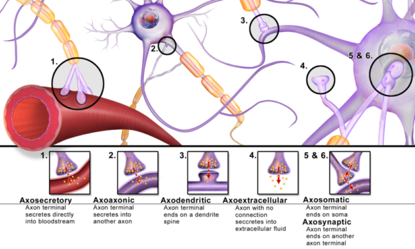
Synaptic transmission sites found in the brain, Blausen/Wikimedia Commons
Synapse structure
Synapse structure is explained as separate compartments. There is a small area between the neuron axon terminal and the cell receiving the neurotransmitter named the synaptic cleft. The part of the axon terminal that releases neurotransmitter is called the presynaptic compartment. The cell structure on the other side of the synaptic cleft where neurotransmitter binds is named the postsynaptic compartment. Each of these parts of the synapse structure possesses a different physiologic function.
Studies of the past 10 years discovered that many axodendritic synapses of the brain have two more functional compartments in their synapse structure. In addition to the two neurons involved, an astrocyte and a microglia participate in the signaling process.
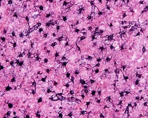
Photomicrograph of protoplasmic astrocytes tiling brain tissue, Jose Luis Calvo/Shutterstock.com
The astrocyte recovers released neurotransmitter from the synaptic cleft by pumping it into its cytoplasm for metabolic processing before sending it back to the presynaptic neuron. The microglia checks to make sure the neuron compartments are in good working order. If there is a problem, microglia works to correct it. Microglia can remove or repair synapses as the need arises.
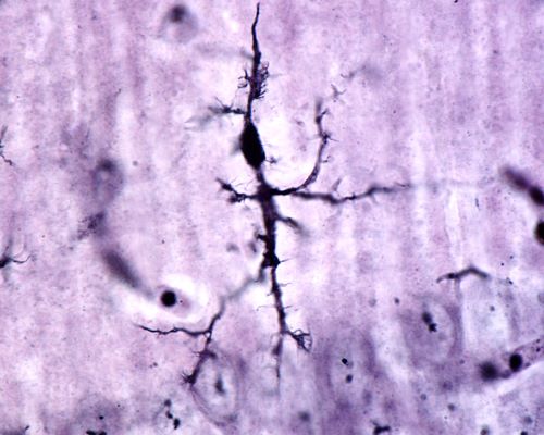
Brain microglia in its neuron monitoring form, Jose Luis Calvo/Shutterstock.com
Neurotransmitter release
There are many different types of neurotransmitters used in the brain, but the mechanism for their release from presynaptic compartments is similar. Neurotransmitter is made by cellular metabolic enzymes in the neuron cell body and is packaged there into vesicles. The vesicles are transported along the internal fibrous tracts through the axon to the presynaptic compartment.
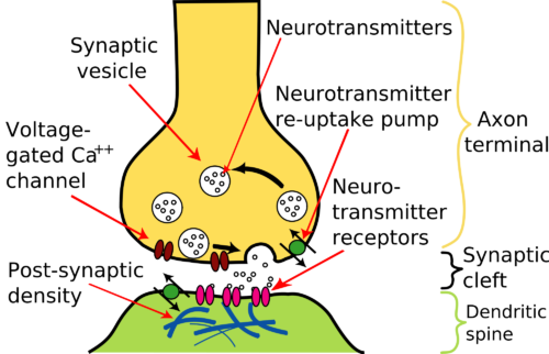
Basic elements of a brain synapse, Nrets/WikiMedia Commons
When action potentials arrive at the axon terminals, they cause the membrane to depolarize. The depolarization affects voltage-sensitive calcium ion [Ca++] channels in the membrane causing them to open. In adult brain the concentration of Ca++ is higher in the extracellular fluid surrounding neuron terminals than inside the neuron. When Ca++ channels open, Ca++ diffuses into the neuron terminal.
The presence of a high level of Ca++ inside the neuron sets in motion a series of molecular processes. This series of events moves the vesicles from their storage area in the axon terminal to the membrane nearest the synaptic cleft. The membranes of the vesicles then fuse with that of the axon terminal such that their neurotransmitter is released into the synaptic cleft.
Synaptic cleft
Neurons usually fire actions potentials as a patterned series. The pattern itself establishes the quality of the message. If neurotransmitter released by each arriving action potential is not removed quickly, the synaptic cleft rapidly becomes saturated making further action potentials ineffective.
Most neurons depend upon neurotransmitter uptake pumps called transporters to remove it from the synaptic cleft. Often most of the neurotransmitter transporters are on the membrane of surrounding astrocyte cells. More than 20 recognized proteins are included in the transporter group. Each type of neurotransmitter uses its own subset of transporter proteins. Neurons releasing the neurotransmitter acetylcholine are an exception to this process. Acetylcholine is inactivated in the synaptic cleft by the enzyme acetylcholinesterase.
Acetylcholinesterase is located on the membrane of the postsynaptic compartment. The enzyme splits acetylcholine into choline and acetate. Choline is then transported back into the presynaptic compartment where it is resynthesized into acetylcholine and repackaged. This approach removes acetylcholine from the synaptic cleft faster than is possible using reuptake membrane pumps.
Neuron dendrites
The majority of postsynaptic compartments on brain neurons are located at structures called dendritic spines on neuron dendrites. Neuron dendrites are branched like small trees and each branch may contain thousands of dendritic spines.
In this image of the cell soma (cell body) and dendrites of a hippocampus neuron notice the tiny protrusions from the dendrite surface. These are the dendritic spines where synapses occur.
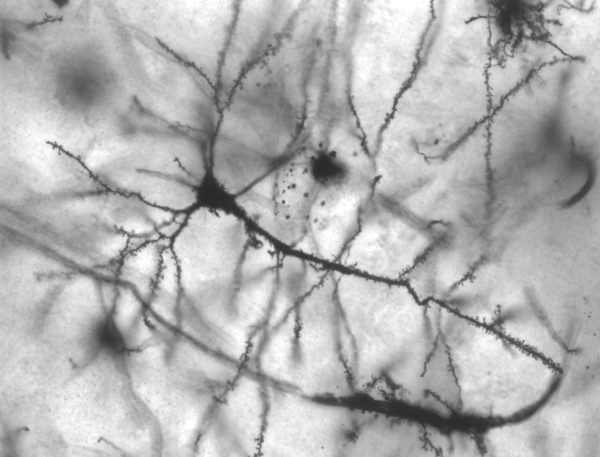
Pyramidal hippocampus neuron showing dendritic spines/MethoxyRoxy/Wikimedia Commons
The main feature of postsynaptic compartments of brain neurons is the postsynaptic density (labeled on the image above) of the plasma membrane of the dendritic spines. The postsynaptic density is a thickening of the plasma membrane that contains a large collection of highly mobile proteins able to respond to neurotransmitter. The diversity and size of this protein population is governed by the pace of neurotransmitter release from the presynaptic compartment.
Some proteins of the postsynaptic density are adhesion molecules that hold the two compartments together, some are enzymes and many are neurotransmitter receptors. Some neurotransmitter receptors open ion channels. These ion channels allow the flow of Ca++, chloride [Cl– ], sodium [Na+] or potassium [K+] into the postsynaptic compartment.
Other neurotransmitter receptors are proteins running through postsynaptic density membrane. The transmembrane receptors alter their shape when neurotransmitter binds. The modification of the receptor’s shape on the inside of the cell sets in motion many intracellular molecular pathways leading to long-term changes in the postsynaptic compartment.
The ion channel receptors for neurotransmitter may cause electrical depolarization of the membrane of the postsynaptic compartment initiating action potentials along the dendrites toward the cell body. Alternately, ion channel receptors may hyperpolarize the membrane and thereby interfere with the production of action potentials along the dendrite.
Positioning of brain synapses combined with the various types of receptors for many different neurotransmitters permits complex synaptic transmission within brain tissue.
For a more in depth review of how brain cells connect, share and disengage to manage the body’s internal and external environment you may be interested in reading my book “Inside the Closed World of the Brain.” For a free look inside this book click here.
If you think this description of brain neuron synapses is helpful, please share it with your fellow students or send it to your favorite social media by clicking one of the buttons shown.
Further Reading:
Neurons and Brain’s Other 90% of Cells

Margaret Thompson Reece PhD, physiologist, former Senior Scientist and Laboratory Director at academic medical centers in California, New York and Massachusetts is now Manager at Reece Biomedical Consulting LLC.
She taught physiology for over 30 years to undergraduate and graduate students, at two- and four-year colleges, in the classroom and in the research laboratory. Her books “Physiology: Custom-Designed Chemistry”, “Inside the Closed World of the Brain”, and her online course “30-Day Challenge: Craft Your Plan for Learning Physiology”, and “Busy Student’s Anatomy & Physiology Study Journal” are created for those planning a career in healthcare. More about her books is available at https://www.amazon.com/author/margaretreece. You may contact Dr. Reece at DrReece@MedicalScienceNavigator.com, or on LinkedIn.
Dr. Reece offers a free 30 minute “how-to-get-started” phone conference to students struggling with human anatomy and physiology. Schedule an appointment by email at DrReece@MedicalScienceNavigator.com.