
Brain capillaries
Brain capillaries prevent 98% of small molecules and almost all large molecules carried in blood from entering the brain. Transport of blood-carried molecules, large and small, into the brain is tightly controlled by an unusual arrangement at brain capillaries called the blood brain barrier. The concentration of water, hormones, amino acids, neurotransmitters and other metabolites fluctuate in blood to a great degree. Such fluctuations in brain tissue would cause unacceptable instability in neural activity.
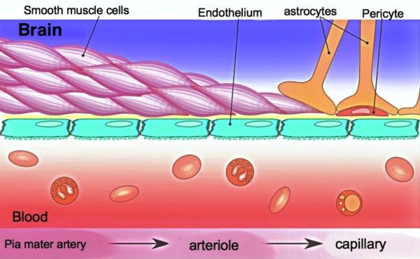
Brain Capillaries are reinforced by pericytes and astrocytes, Armin Kubelbeck/Wikimedia Commons
The blood brain barrier is created by tight lacing together of capillary endothelial cells by protein formations called tight junctions and adherens junctions. The junctions create a situation where all molecules must be transported from the capillaries through, instead of around, the endothelial cells.
An additional layer of physical and functional support for the blood brain barrier comes from pericytes, cells that wrap around the capillaries. Addition strength is added to the blood brain barrier by projections of astrocyte glial cells that make direct contact with capillary endothelial cells. The three cell types virtually enclose the entire system of brain capillaries.
Blood brain barrier transport
The blood brain barrier transport system is composed of multiple membrane transporters. Oxygen and carbon dioxide are lipid soluble and pass easily through the cell membranes of the barrier. Some other small lipid molecules also pass through the blood brain barrier, but most are subsequently transported back to the vascular system by cell efflux pumps.
It has been estimated that 10-15% of all proteins in the brain’s vascular system are transporters to move substances across the capillary endothelial cells. Glucose, the main energy substrate of the brain, is transported into brain by a transporter named the GLUT1 capillary endothelial cell transporter. There are also transporters for the amino acids needed to synthesize the neurotransmitters norepinephrine, dopamine, and serotonin.
Entry of water and other molecules necessary for brain health occurs primarily through the capillaries of the choroid plexus in the brain’s central chambers, the ventricles. Each of the four ventricles has a choroid plexus that produces cerebrospinal fluid. The choroid plexus selectively acquires blood molecules essential for brain health and uses them to create the cerebrospinal fluid. The cerebrospinal fluid circulates through and around the brain and spinal cord.
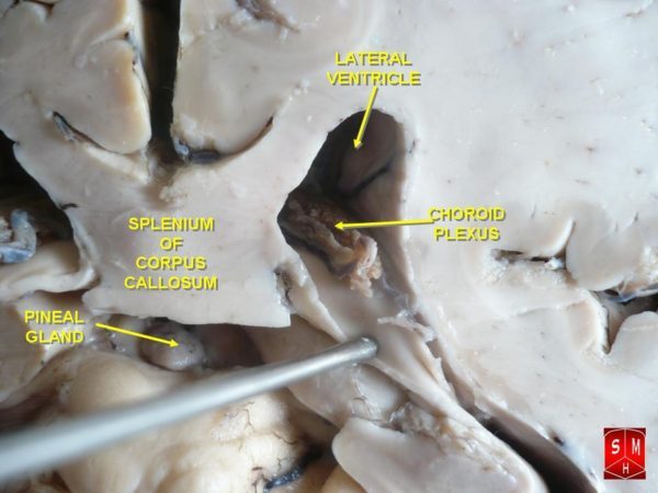
Choroid Plexus of the lateral ventricle of the human brain, Anatomist90/Wikimedia Commons
Nutrients are supplied to the interstitial fluid surrounding neurons and glia by cerebrospinal fluid. In turn, metabolic waste from the interstitial fluid is delivered into the cerebrospinal fluid and ultimately returned to the venous drainage of the brain.
Brain membranes
Brain membranes, the meninges, are another component of the brain’s barrier system. Between brain’s neural tissue and its blood vessels there is a fluid filled space named the Virchow-Robin Space created by brain membranes. There is a layer of thin membrane that covers the surface of the brain called pia mater. Notice its location in the image below. The name comes from Medieval Latin and it translates to ‘tender mother’. Pia mater also dips into the substance of the brain and forms a sheath around the brain’s arteries and arterioles.
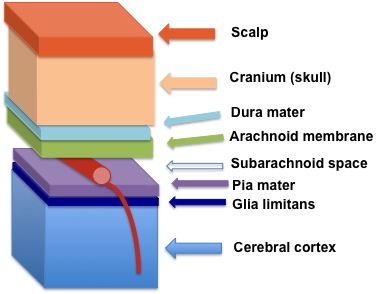
Brain meninges as they appear in layers over the cerebral cortex, , Katie Ahlers/Wikimedia Commons
Large arteries supplying the brain are branches of the internal carotid arteries of the neck and the vertebral arteries. The pia mater sheath around the larger arteries entering the brain through the subarachnoid space is continuous with that of the smaller penetrating arteries and arterioles of brain tissue.
The space between the pia mater sheath and arteries is called the Virchow-Robin space. The Virchow-Robin space is filled with interstitial fluid and it has important connections with the lymphatic drainage of the head. In normal brain, any pathogen or stray white blood cell that manages to escape the arteries or arterioles of the brain is quickly removed by the Virchow-Robin space. The Virchow-Robin brain membrane space ends where blood capillaries and the blood brain barrier begin.
Brain immune system
The brain immune system is separate from the blood vessel inflammation response elsewhere in the body. What is it about how a brain works that it is advantageous for it to bypass the primary defense system used by the rest of the body, leukocyte respondents? When damage occurs within the healthy brain, local microglia is the first to react. Microglia kills pathogens, clears away the destruction and repairs the damage without edema and extra heat in the tissue.
Managing a classic peripheral immune response is troublesome for the brain, because there is very little room for expansion within the skull. The immune response outside the brain to injury and pathogens includes swelling (edema), heat in the tissue, and loss of function in the area affected. There is increased blood flow, increased capillary permeability, and increased macrophage, T-Cell and B-Cell infiltration of the tissue.
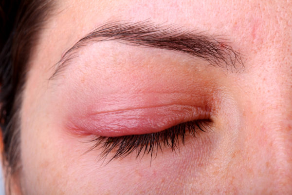
Woman’s eyelid showing red swollen tissue of an immune response, Zenakoroleva/Shutterstock.com
However, even under normal circumstances a small number of T-Cells routinely enter the cerebrospinal fluid at the choroid plexuses of the ventricles. They appear to be a standby support system for the microglia. Microglia can present antigen to surveillance T-Cells when damage is more than it can handle on its own. Recruiting a T-Cell response in extreme situations changes the permeability of the blood brain barrier to the immune system that functions primarily outside the brain.
Further reading
Quality Control of Brain’s Watery Environment
Stem Cells: An Evolving Definition
Do you have questions?
Send me an email with your questions at DrReece@MedicalScienceNavigator.com, or put them in the comment box. I always read my mail and answer it. Please share this article with your fellow students taking anatomy and physiology by clicking your favorite social media button.

Margaret Thompson Reece PhD, physiologist, former Senior Scientist and Laboratory Director at academic medical centers in California, New York and Massachusetts is now Manager at Reece Biomedical Consulting LLC.
She taught physiology for over 30 years to undergraduate and graduate students, at two- and four-year colleges, in the classroom and in the research laboratory. Her books “Physiology: Custom-Designed Chemistry”, “Inside the Closed World of the Brain”, and her online course “30-Day Challenge: Craft Your Plan for Learning Physiology”, and “Busy Student’s Anatomy & Physiology Study Journal” are created for those planning a career in healthcare. More about her books is available at https://www.amazon.com/author/margaretreece. You may contact Dr. Reece at DrReece@MedicalScienceNavigator.com, or on LinkedIn.
Dr. Reece offers a free 30 minute “how-to-get-started” phone conference to students struggling with human anatomy and physiology. Schedule an appointment by email at DrReece@MedicalScienceNavigator.com.