
Begin learning the GI tract with its enteric nervous system
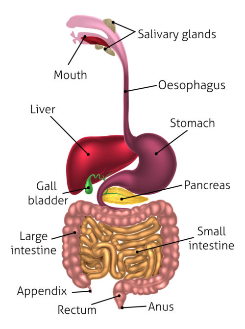
Gastrointestinal tract, Cristos Georghioous/Shutterstock.com
Before starting a discussing of neural control of the gastrointestinal tract, a quick review of the general organization of the human nervous system is necessary. The nervous system has three parts to its organization. First, there is a set of neurons that sense what is happening in their local environment and relay that information. Second, there is a receiver for the sensory information that interprets the data, the central nervous system (CNS). The CNS is composed of the brain and the spinal cord. Third, there are the motor neurons of the brain that respond with signals that cause muscle contraction.
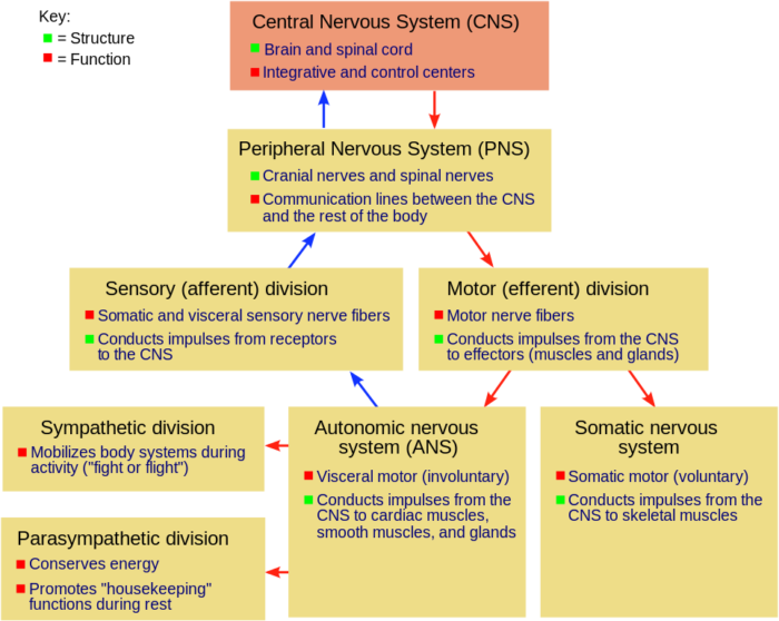
Parts of the nervous system, Fuzzform at English Wikipedia/Wikimedia commons
Three types of muscle tissue are found in the human body, heart muscle, skeletal muscle and smooth muscle. Heart muscle generates its own electrical activity. The brain’s motor neurons for skeletal muscle are under conscious control. They are called the somatic motor neurons. The brain’s motor neurons for smooth muscle operate automatically and are named the autonomic nervous system.
Gastrointestinal tract muscle
Skeletal muscles associated with chewing and swallowing are controlled by somatic motor neurons. However, most of the gastrointestinal tract is composed of smooth muscle, which is regulated by the autonomic nervous system. It is usually stated that there are two opposing divisions of the autonomic nervous system, the sympathetic and parasympathetic. However, to explain gastrointestinal physiology a third division of the autonomic nervous system must be included, that is the enteric nervous system.
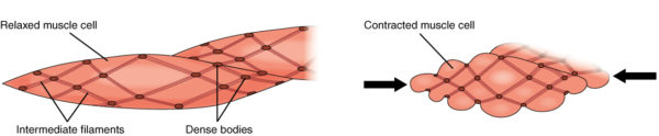
How smooth muscle shortens, OpenStax College/Wikimedia Commons
The enteric nervous system resides completely within the layers of the gastrointestinal tract, esophagus to anus. The enteric nervous system controls movement for food through the tract. Its activity may be hastened or slowed by activity of the sympathetic or parasympathetic neurons, but the basic rhythm of enteric neuron firing occurs without central nervous system management.
Enteric nervous system origin
The enteric nervous system is a mesh-like network of neurons and glial cells that arise in the embryo from neural crest cells. All four major classes of neurons are present. There are motor neurons, sensory neurons, interneurons and ganglionic neurons. In the brain, clusters of neuron cell bodies are called nuclei. Outside the central nervous system, clusters of neuron cell bodies are called ganglion. The ganglion of the intestine are volume sensors.
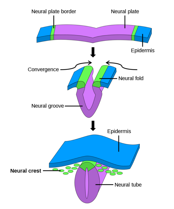
Neural crest formation in an embryo, Public Domain/Wikimedia Commons
Brain neurons and brain glial cells also derive from neural crest cells of the embryo. There are many similarities in the embryonic lineage and functional relationships between glial cells and neurons of the gastrointestinal tract and brain. Glial cells of the gut include Myelinating Schwann cells and Non-myelinating Schwann cells. Similar myelinating cells in the brain are called oligodendrocytes.
In addition to myelination of neuron axons, brain and gut glial cells performs many functions in support of neurons including regulation of neuron synapses. A portion of the Non-Myelinating Schwann cells resemble and perform like brain astrocytes, another type of glial cell. The circuits of the enteric neurons control smooth muscle contraction, local blood flow, mucosa transport and secretion and they modulate immune function.
Gastrointestinal tract tissue layers
The tubular gastrointestinal tract has several structural layers. The innermost layer is the mucosa surrounding the lumen. The mucosa comes in direct contact with digested food. It is made up of 3 layers, which are the epithelium, the lamina propria and the muscularis mucosa. The inner epithelial layer is where most digestive, absorptive and secretory processes occur. It is the epithelial layer that varies most along the length of the tract. It changes by region to deal with the different work of each section of the tract in breaking down food particles, absorbing nutrients and eliminating waste.
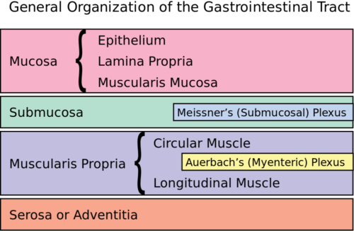
Layers of the GI tract, Hazmat2/Wikimedia Commons
Beneath the mucosa is a layer of connective tissue, the submucosa. One part of the enteric nervous system, Meissner’s Plexus which is also called the Submucosal Plexus, lies within the submucosa.
Two outer layers of smooth muscle running the length of the tract comprise the muscularis propria. The outermost muscle layer extends longitudinally along the tubular surface. The inner muscle layer encircles the tract. Between the layers of smooth muscle lies the second net-like layer of enteric neurons, Auerbach’s Plexus also referred to as the Myenteric Plexus. This positioning permits coordinated contraction of the layered muscles to move food through the gastrointestinal tracts sequential stations specialized for breakdown of food particles and absorption of nutrients.
The outer layer of the tube, the serosa or adventitia is connective tissue that produces lubricant to reduce friction during digestive movements.
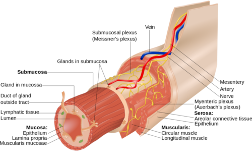
Epithelial, muscle, nerve and blood vessels of the gastrointestinal tract, Goranteken/Shutterstock.com
Regulation of enteric neurons
Enteric neurons’ control of the gastrointestinal tract is influenced by the central nervous system via the autonomic neurons. Neurons of brain stem nuclei direct output of the sympathetic and parasympathetic autonomic neurons that synapse on the enteric neurons of the gastrointestinal tract. The digestive system can function without this regulation, but it is more responsive to environmental circumstances with it.
Parasympathetic effects
Parasympathetic activity increases muscle tone, motility, gastric acid secretion and firing of enteric neurons. Parasympathetic innervation to the stomach, small intestine and proximal colon is supplied by the vagus nerve, which is Cranial Nerve X.
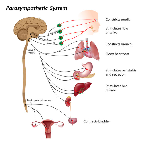
Parasympathetic nerves and organs they innervate, Alila Medical Media/Shutterstock.com
Cell bodies of the motor neurons contributing to the vagus nerve lie in a region of the brain called the dorsal motor nucleus of the vagus or DMV. Vagus motor neurons fire action potentials spontaneously at a low frequency. They receive input from other parts of the brain including the hypothalamus and amygdala that causes their spontaneous rate to increase or decrease. Because the DMV is a circumventricular organ in the fourth ventricle, it lacks a blood brain barrier. Without a capillary barrier, blood molecules also affect firing rate of the vagus motor neurons.
The vagus is a mixed sensory and motor nerve. Remember a nerve is a bundle of many neuron axons. The vagus nerve can be 70% to 80% sensory neuron axons. Sensory neurons of the gastrointestinal tract are ganglion cells. Axons of the ganglion cells travel in the vagus nerve and relay their information to multiple brain nuclei. They signal about gut distention, pressure, pain and the osmotic character of digestive fluids. All such incoming sensory information is processed by various brain areas and relayed to the vagus motor neurons of the DMV.
Sympathetic effects
Sympathetic nervous system activity inhibits GI tract functions. The stomach, small intestine and proximal large intestine are innervated by both parasympathetic and opposing sympathetic neurons. Dense sympathetic synapses populate gastrointestinal tract sphincters producing constriction.
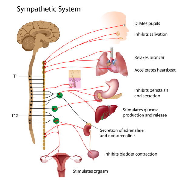
Sympathetic nervous system and the organs they innervate, Alila Medical Media/Shutterstock.com
However, sympathetic innervation of the circular and longitudinal muscle is sparse. Instead this system of neurons slows the firing rate of the enteric neurons of the myenteric and submucosal plexus by acting to block neurotransmitter at enteric neuron synapses. Sympathetic nerves are mixed function. About 50% of the axons of sympathetic nerves are sensory and relay information to brain nuclei.
Balancing autonomic nervous system activity
Both parasympathetic and sympathetic neurons fire continuously. A change in firing rate of one is always accompanied by an opposite change in firing rate by the other. An increase in parasympathetic activity is always matched by a decrease in sympathetic activity. An increase in sympathetic activity is always matched by a decrease in sympathetic activity. It is the net analysis of incoming sensory information to brain nuclei that determines which system’s output will dominate at any time.
Because the GI tract and enteric nervous system are so greatly influenced by the sympathetic and parasympathetic systems, its optimal function is a lower priority in some situations. During exercise or fight-or-flight situations sympathetic activity dominates and food digestion slows. During restful times parasympathetic activity dominates and digestion proceeds normally.
Summary of GI physiology
- The enteric nervous system manages movement of ingested material from esophagus to anus.
- The enteric nervous system is a division of the autonomic nervous system, because it operates subconsciously and triggers contraction of smooth muscle cells.
- Embryonic origin, differentiation and function of enteric neurons and glial cells are like that of parallel cell populations in the brain.
- Parasympathetic neurons are supportive of enteric nervous system processing of food and nutrient uptake into blood.
- The mixed function parasympathetic vagus nerve provides a path for sensory information about the digestive process to reach brain nuclei.
- Sympathetic neurons inhibit the neurons of the enteric nervous system and slow food processing.
- Cortical brain neurons respond to sensory information from the gastrointestinal tract by signaling to the brain stem center controlling parasympathetic motor output.
- In times of exercise or emergency, the sympathetic nervous system dominates, and resources devoted to digestion are diverted.
Further reading
Human Digestion: Main & Accessory Organs
Gastrointestinal Hormones for Digestion & Fasting
Neurons: Where does their electricity come from?
Do you have questions?
Please put your questions in the comment box or contact Dr. Reece directly at DrReece@MedicalScienceNavigator.com, or on Pinterest, or on LinkedIn.

Margaret Thompson Reece PhD, Physiologist, Medical Research Scientist and Laboratory Director during over 20 years in OB/Gyn clinical departments at academic medical centers in California, New York and Massachusetts, is now Manager at Reece Biomedical Consulting LLC.
Asna educator, she taught physiology for over 30 years to undergraduate and graduate students, at two- and four-year colleges and universities, in the classroom and in the research laboratory.
Experience taught here that the best way for premed students to get the B+ or better grade in A&P that is required for acceptance to medical training, the first time through the course, is to describe body processes on exams in terms of neural, cardiovascular, and endocrine involvement.
Her books help A&P students struggling to master the chemistry of human physiology. They are “Physiology: Custom-Designed Chemistry” and “Inside the Closed World of the Brain.” More about her books is available at https://www.amazon.com/author/margaretreece.
For those preferring video training, she offers an online course “30-Day Challenge: Craft Your Plan for Learning Physiology”
Dr. Reece offers a free 30 minute “how-to-get-started” phone conference to students struggling with human anatomy and physiology. Schedule an appointment by email at DrReece@MedicalScienceNavigator.com.
.