
Only 4 human tissue types, why are they so hard to see?
Most A&P courses teach tissue types early in the laboratory set of lessons. While it makes sense to start lab with the smallest components of the body and work up from there, tissue lab is very hard for beginning students and for their instructors to explain. This article will help you get started toward an understanding what you are seeing under the microscope or in photomicrographs for your lab practical exam.
Lab practical exam
One segment of anatomy lab practical exams that frustrates most students, and many instructors as well, is recognizing types of human tissues under the microscope. For many students this is their first time using a microscope or viewing photomicrographs, pictures of tissue sections captured with a microscope.
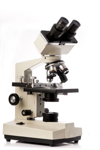
Standard upright light microscope, Brian Maudsley/Shutterstock.com
If your instructor wants you to use a microscope in lab, you may find that just locating the tissue on the slide and bringing it into focus are major challenges. Then once you can see the tissue, you may still be at a loss about which features are important.
Even after you get finding the tissue section and focusing the microscope under control, remembering what you saw through in the microscope will be less than precise. The human mind has a limited ability to hold the fine details of an image in memory.
To perform well on the tissue lab practical exam, it is helpful to know ahead of time what you are trying to remember about each tissue type. For simplicity, anatomists divide animal tissues into four types, connective tissue, muscle tissue, nervous tissue, and epithelial tissue based upon their structural features.
At the magnification possible with a standard lab light microscope, usually you will see more than one type of tissue in the visual field. So, the first thing you want to do is to decide which tissue types are present.
Tissue types organize for function
The tissue types present and their organization in a histologic section indicate the organ source of the tissue and the organ’s function.
In the body, the tissue types and sub-types are mixed to support function. For example, it is functionally an advantage for nerves, arteries, and veins to travel tougher through the body in bundles.
The bundles are tied together by fascia. Fascia is a sheet of connective tissue, primarily composed of collagen, that encloses and stabilizes bundled tissues.
Here is a photomicrograph of a vascular bundle travelling through adipose tissue.
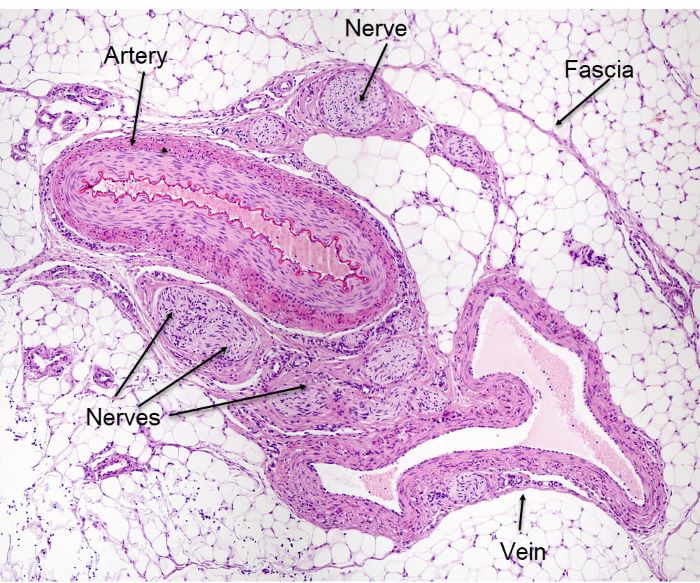
Vascular bundle in fat tissue, Jose Luis Calvo, Shutterstock.com
Tissues within this vascular bundle include smooth muscle and elastic cartilage of a thick-walled artery, smooth muscle of a large vein, bundles of neuron axons shown as cross sectioned nerves, endothelia tissue lining the cavity of the artery and vein.
The entire vascular bundle lies in a connective tissue named adipose (fat). Blood capillaries supplying the fat are composed entirely of endothelia cells, a subtype of epithelium.
As you later study the anatomic pathways of arteries, veins, and nerves keep the structure of vascular bundles in mind. Because arteries, veins, and nerves travel together, the components of the bundle in each anatomic region will have similar names.
Tissue sub-types
Tissue identification is complex because each of the four basic tissue types has a variety of sub-types. Tissue sub-types are identified by their shape and their ability to take up various dyes. Each histologic section usually contains more than one of the many possible combinations of tissue sub-types.
During a lab practical exam, it is usually required that you distinguish which organ an arrangement of the tissue types represents. To do this, you must look for clues about the identity of the organ by observing the sub-types of tissues present. Think about how each of the sub types may support an organ’s function.
The image below shows a sub-type of nervous tissue and a sub-type of muscle tissue. This image is also in the website banner at the top of this page.
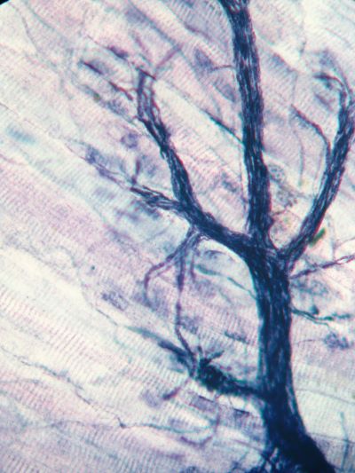
Skeletal muscle with and associated motor neuron, Jubal Harshaw/Shutterstock.com
If you look closely, you will see tube shaped cells with stripes. The stripes are characteristic of muscle cells. The long tube shapes running in parallel identify it as skeletal muscle.
The dark blue string-like structure overlaying the striped tubes is nervous tissue, in this case a motor neuron. So, there are two sub-types of tissue in this photomicrograph.
Notice how the motor neuron branches at its end to control individual muscle cells. This combination of neuron and skeletal muscle is called a skeletal muscle motor unit.
Below is another example of a combination of tissues used to form a body organ.
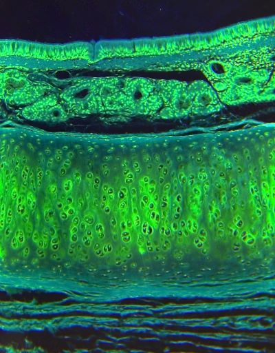
Cross section of a trachea with cartilage and cilia, Jubal Harshaw/Shutterstock.com
This is a piece of a cross section of a trachea that includes connective tissue, cartilage, and epithelial cilia covering the organs air surface. Beneath the cilia and above the cartilage are glands that are also composed of epithelial cells.
The cartilage holds the trachea open so that air can flow to the lungs. The glands are there to secrete mucus to trap unwanted particles in the air before they get to the lungs. The cilia motion drives the mucus toward the mouth to expel it from the body.
Overtime you will come to recognize the patterns of cells you observe with the light microscope. When you are just beginning, list what types of tissue layers you observe and then think about how those tissue may help a body organ achieve a goal.
There is a great online resource to help you become very good at identifying human tissue specimens.
Best tissue identification resource
The best resource I have found for figuring out what I am looking at through a microscope is a group of videos posted on YouTube by a teaching pathologist, Dr. John Minarcik. Even though I have used a microscope in my scientific studies for decades, when I am in doubt about a human tissue, I go ask a pathologist for help.
Watch this video by Dr. Minarcik then look again at the photomicrograph or the cross section of the trachea.
For more videos by Dr. Minarcik about tissues, search YouTube for ‘Shotgun Histology’ or click on the Human Tissue Histology in the ‘Further reading” list below or click here.
A very nice feature of this series of YouTube videos is that you can view them enough times that the images become familiar to you. Familiar images are memorable images. Also, the various parts of each image will make more sense to you.
It is much easier to remember an image when there is logic to the detail. With this kind of help, tissue anatomy lab exam may become your easiest lab practical exam.
Further reading
Memory, Patterns and Themes in A&P
Do you have questions?
Please put your questions in the comment box or send them to me by email at DrReece@MedicalScienceNavigator.com. I read and reply to all comments and email.
If you find this article helpful share it with your fellow students or send it to your favorite social media site by clicking on one of the buttons below.

Margaret Thompson Reece PhD, physiologist, former Senior Scientist and Laboratory Director at academic medical centers in California, New York and Massachusetts is now Manager at Reece Biomedical Consulting LLC.
She taught physiology for over 30 years to undergraduate and graduate students, at two- and four-year colleges, in the classroom and in the research laboratory. Her books “Physiology: Custom-Designed Chemistry”, “Inside the Closed World of the Brain”, and her online course “30-Day Challenge: Craft Your Plan for Learning Physiology”, and “Busy Student’s Anatomy & Physiology Study Journal” are created for those planning a career in healthcare. More about her books is available at https://www.amazon.com/author/margaretreece. You may contact Dr. Reece at DrReece@MedicalScienceNavigator.com, or on LinkedIn.
Dr. Reece offers a free 30 minute “how-to-get-started” phone conference to students struggling with human anatomy and physiology. Schedule an appointment by email at DrReece@MedicalScienceNavigator.com.